01 Summary
This application note demonstrates the highly efficient ex vivo expansion of primary human T cells using Hoxton Pharma’s proprietary bioreactor platform that meets the unique demands of both autologous and allogeneic immunotherapies. The system achieves primary T cell proliferation rates and critical quality attributes that match or exceed traditional high-cost, single-use static culture systems such as the G-Rex® platform, for different donors tested.
02 Background
In the rapidly evolving biopharmaceutical landscape, chimeric antigen receptor (CAR) T-cell therapies have emerged as a paradigm-shifting modality for oncology, delivering unprecedented clinical outcomes in haematologic malignancies. However, the commercial scaling of these therapies remains severely bottlenecked by the technical and economic realities of autologous manufacturing. Because autologous CAR-T cell production relies entirely on patient-specific cell material, manufacturing cannot utilise traditional, large-scale bulk processing. Instead, it demands a “scale-out” architecture where a single, dedicated manufacturing run must be precisely executed for every individual patient.
Ex vivo T-cell expansion is a critical and highly sensitive phase of this scale-out workflow. Traditionally, this process relies on passive, gas-permeable static culture systems like G-Rex® (Wilson Wolf) devices or complex systems such as CliniMACS Prodigy® Instrument (Miltenyi Biotec) (Levine, Bruce L et al., 2016). However, replicating thousands of these individual static vessels across a manufacturing facility dramatically swells the cleanroom footprint and drives up the cost of specialised, single-use consumables. Consequently, the industry faces an urgent need for automated, closed-loop systems capable of delivering high-density, high-quality T-cells within a highly compact operational profile.
To address these critical bottlenecks, this application note introduces Lattice mini, an automated, small-scale platform purpose-built for decentralised manufacturing of human cells, including scale-out primary T-cell expansion. Our system facilitates gas exchange with active fluid dynamics and automation control which reduces manual intervention for culturing tracking and hence operational variability. By transforming cell expansion from a labour-intensive, passive process into an automated, low-interaction and controlled operation with ease of sampling, the Lattice bioreactor system offers a reliable and economically viable path toward scaling autologous and allogeneic cell therapies to meet global patient demand.
03 Objective
This study aims to demonstrate that Lattice can support the ex vivo expansion of primary T cells across multiple donors with high total fold expansion amenable for clinical applications and maintenance of desirable T cell phenotype for effective patient treatment.
04 Materials & Methods
Bioreactor system
Cultivation was conducted in Hoxton Pharma’s proprietary modular bioreactor system (Lattice mini). Each vessel consists of a flexible, gas-permeable membrane operating at a 0.1–0.5 L working volume. Vessels are mounted in a common frame that provides automated agitation via rocking/rotational motion, eliminating the need for high-shear internal mixing and active gassing. Fluid transfers were managed through aseptic ports linked to a common fluid handling system. The vessels were autoclaved and integrity-tested prior to use.
Biological system and process parameters
Primary pan-T cells isolated from 3 donors (StemCellTechnologies, Cat No. 70024) were inoculated into the vessels at a target density of 2e5/mL. The cells were kept in Lattice mini for a total of 5 days. Environmental and mechanical parameters were maintained as follows:
- Target temperature: 37°C
- Acceptable pH range: 6.5–7.5
- Gas exchange range: 0–5% CO2 (passive O2/CO2 diffusion via the gas-permeable membrane)
- Agitation: 6° oscillation amplitude, 5 deg/s² acceleration, and 3 s pause duration
- Feeding strategy: fed-batch; a single top-up was performed following inoculation
- Metabolites/pH and gas monitoring: offline tracking of glucose, glutamine, lactate, and glutamate as well as pH, pCO2 and pO2 was measured on a BioProfile® FLEX2 analyser
- Sterility: culture sterility was verified by microscopic inspection
Experimental design
Following an adherent expansion phase, cells were inoculated in the vessels at 2e5/mL at Day 0 of reactor culture. Activation beads were kept throughout the culture until harvest. Equal split ratios were used across the vessel types.
- Counting and flow timepoints: Days 1, 2, 4 and 5
- Working volumes: Lattice mini, 250–500 mL; G-Rex® 10M, 100 mL
- Culture media: TexMACS media (Miltenyi Biotec, Cat No. 130-097-196) supplemented with 10% Fetal Bovine Serum (FBS) and 200 IU/mL of human recombinant IL-2 (Biolegend, Cat No. 589104)
- T cell activation method: Dynabeads™ Human T-Activator CD3/CD28 for T Cell Expansion and Activation (ThermoFisher, Cat No. 11132D)
05 Results
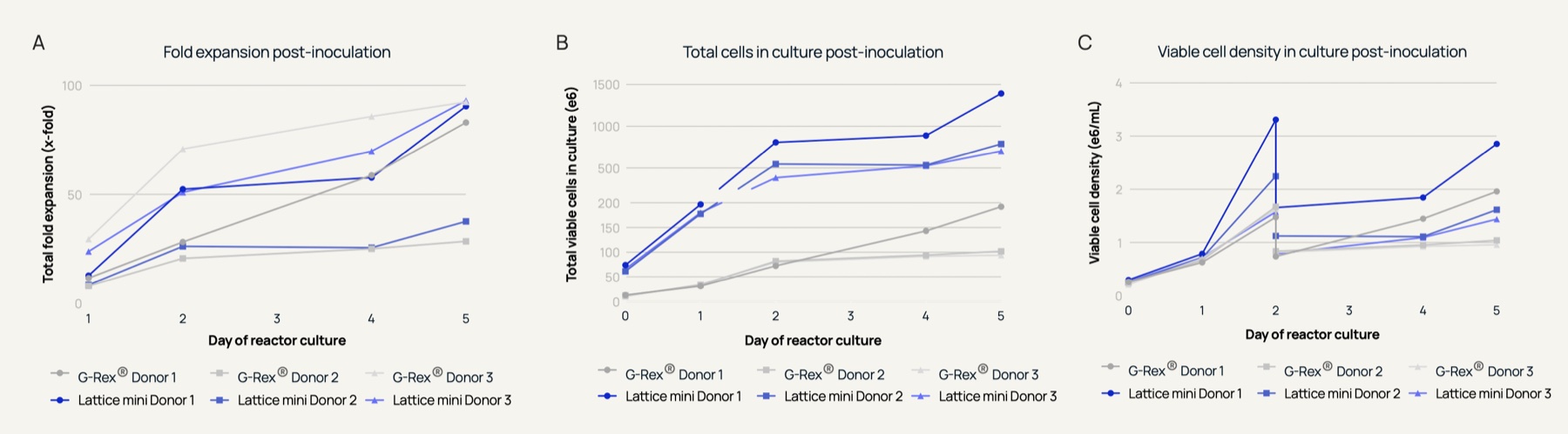
Lattice mini achieves comparable fold expansion to G-Rex®, with similar morphology and metabolic trends
T cells expanded in Lattice mini and G-Rex® show similar phenotype
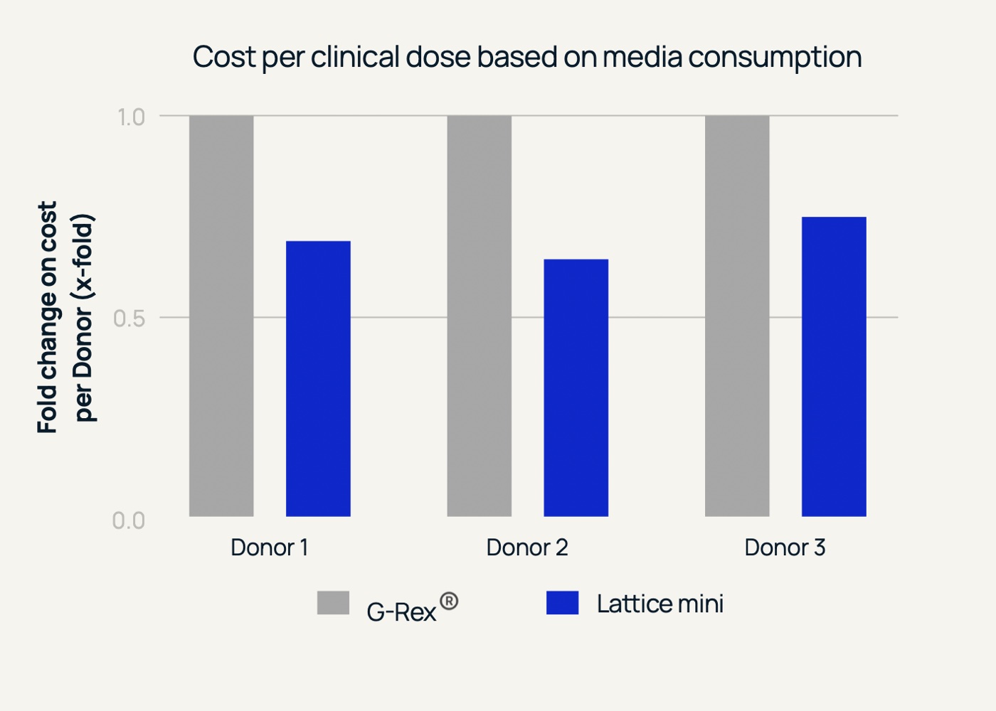
Production of T cells in Lattice mini requires lower volume of culture media than G-Rex®
06 Discussion
The commercial success and accessibility of autologous CAR-T cell therapies depend heavily on manufacturing platforms that can reduce costs while maintaining or enhancing cell quality. This study evaluated the performance of the proprietary Lattice mini bioreactor against one of the industry gold standards, the G-Rex® bioreactor, across three distinct human donors. By focusing on primary T cell culture and expansion without transduction, this evaluation isolates the structural and operational impacts of the Lattice vessel design on cell fitness, growth yield, and economic efficiency.
The primary benchmark for any cellular manufacturing platform is its capability to expand primary cells to therapeutic quantities within a tight clinical window. As detailed in Figure 1, the Lattice mini bioreactor demonstrates growth kinetics that are fully comparable to, and in several instances exceed, the G-Rex® gold standard as evidenced by the total fold expansion metric. Another key finding of this study is the system’s ability to support high-density cultures, surpassing viable cell densities of 3e6 cells/mL during the active growth phase depending on the donor.
Furthermore, the expansion capability within the Lattice vessel shows excellent scaling efficiency. Depending on individual donor characteristics, the platform demonstrated that an initial target seed stock of 50e6 (actual 74e6) cells could be successfully expanded to yield over 1e9 total viable cells in the 5-day period post-inoculation while utilising a total of only 500 mL of culture media. This rapid, high-density proliferation satisfies the baseline yield requirements for manufacturing single clinical doses, making it an incredibly robust alternative for cell therapy developers.
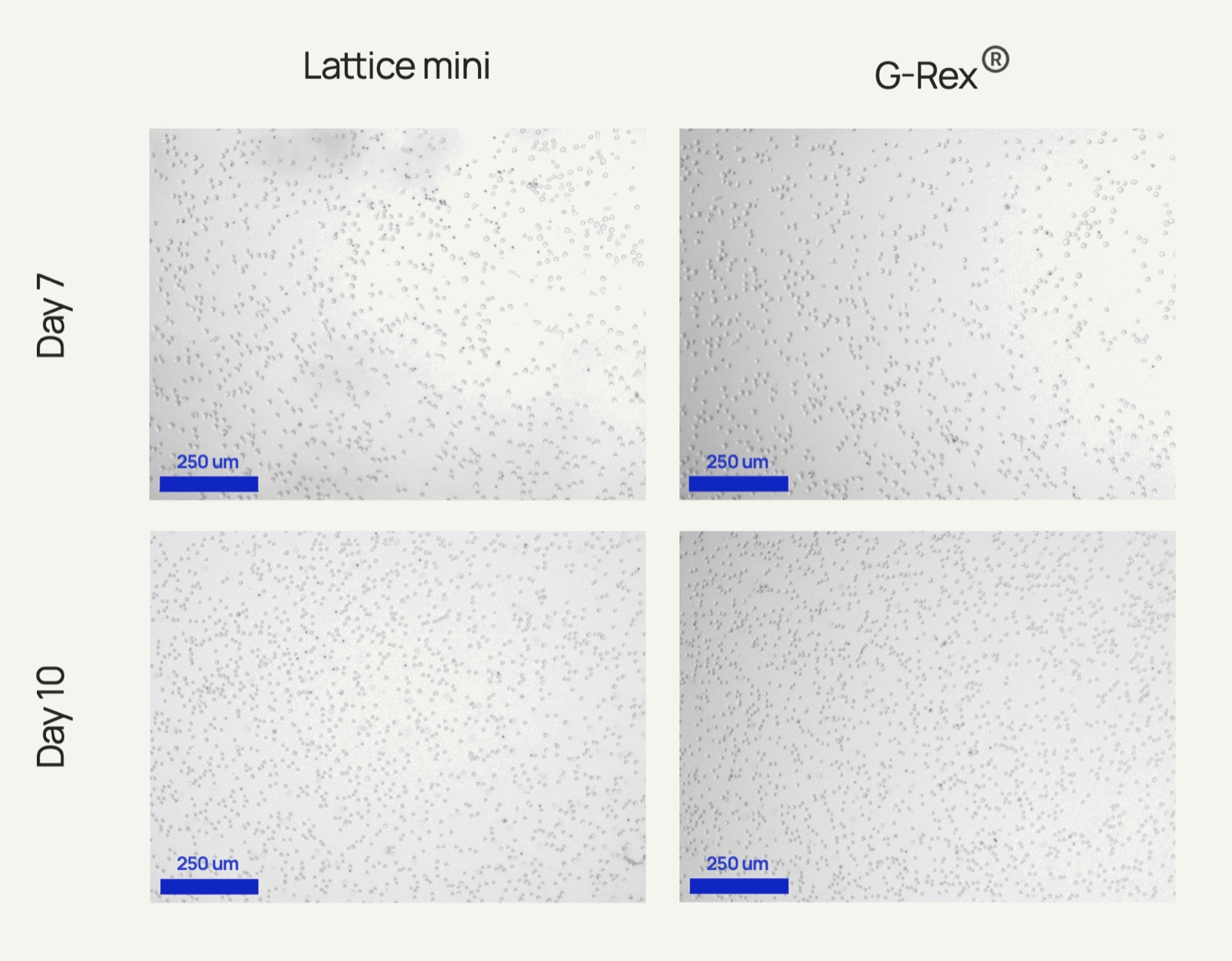
Visual inspection of the expanded T cells provides essential context regarding culture homogeneity and overall health. Microscopic analysis captured in Figure 2 reveals the cellular morphology of the primary T cells on Day 7 and Day 10 across all three donors. In both the G-Rex® and Lattice mini vessels, the cells maintain a healthy, spherical, non-adherent appearance characteristic of robust primary T cell cultures. Crucially, the cells in the Lattice vessel exhibit a highly uniform distribution without forming aggregates that can lead to localised nutrient starvation or necrotic cores. This structural uniformity validates that the internal architecture and rocking of the Lattice bioreactor delivers a well-balanced physical microenvironment. It ensures that even at elevated cell densities, every cell retains good surface exposure to the surrounding nutrient medium.
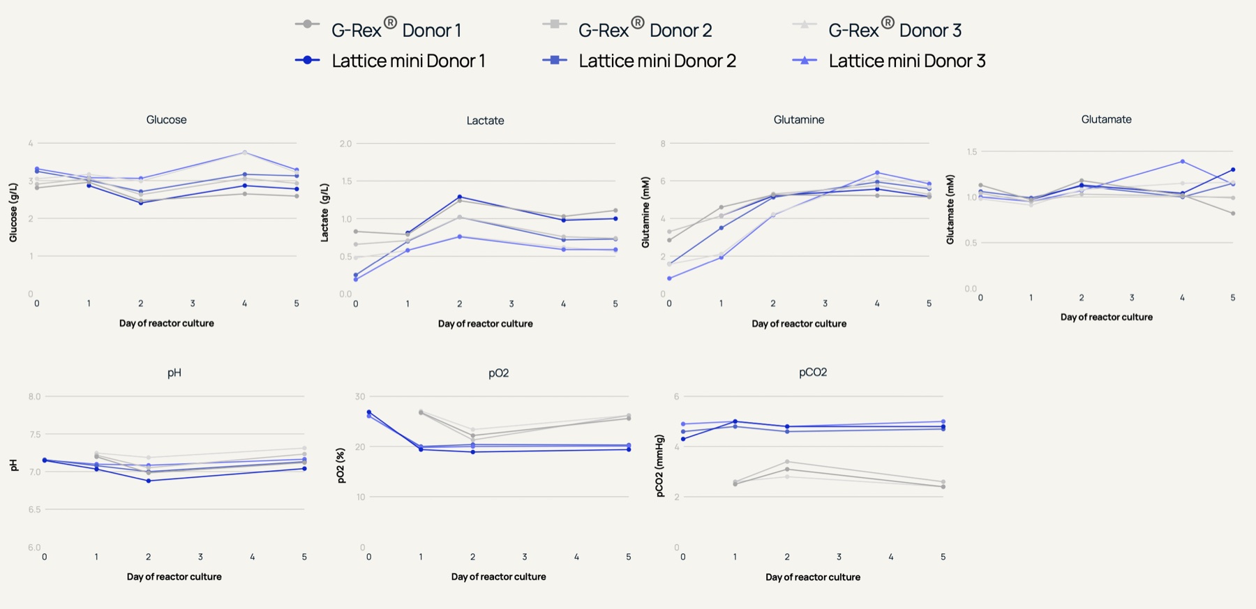
Besides cell morphology, monitoring metabolic trends is a reliable method for assessing cellular stress and ensuring process consistency. As illustrated by the metabolic profiles in Figure 3, the consumption of glucose and glutamine, alongside the production of lactate and glutamate, followed similar trends between the Lattice and G-Rex® vessels for each respective donor. This parallel behaviour indicates that the physical configuration of the Lattice bioreactor does not alter standard primary T cell metabolism or induce metabolic stress. Environmental parameters further underscore the stability of the Lattice platform. The pH, dissolved oxygen (pO2), and carbon dioxide (pCO2) levels remained steady and tightly controlled throughout the culture duration. The efficient gas exchange and stability shown in these profiles confirm that the Lattice platform successfully prevents toxic byproduct accumulation while meeting the oxygen demands of dividing primary T cells.
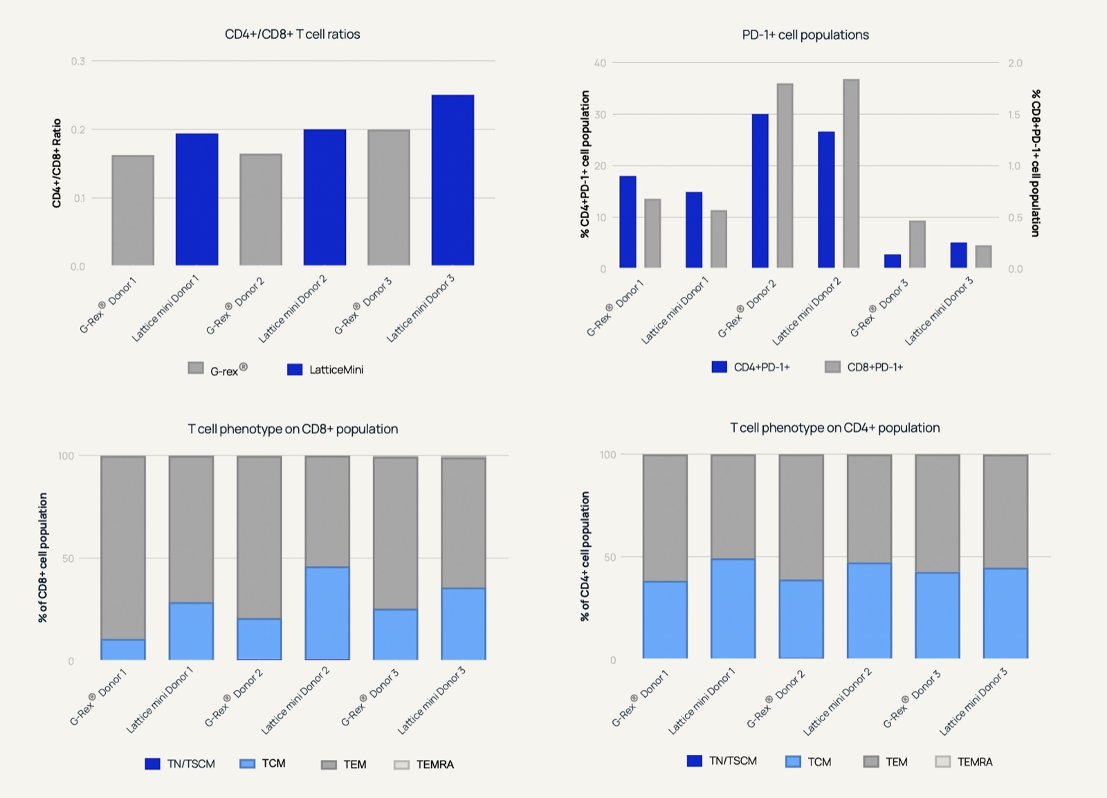
While cell quantity is vital, the phenotypic composition of the final product determines clinical efficacy. Flow cytometry data in Figure 4 highlights an advantageous shift in cell composition within the Lattice bioreactor. For the specific protocol used, primary T cells expanded in the Lattice vessel displayed a higher proportion of the T central memory (TCM) phenotype and a lower percentage of the T effector memory (TEM) phenotype compared to those grown in the G-Rex® system across both CD4+/CD8+ populations. Exhaustion markers like PD-1 remained comparable between both systems and mostly differed on a donor-specific basis.
In a clinical context for T cell therapy, maintaining a less-differentiated, memory-rich phenotype (TCM and TSCM) is correlated with superior in vivo persistence, sustained proliferation, and better long-term patient outcomes post-infusion (López-Cantillo, Gina et al., 2022). Expansion of primary T cells in Lattice clearly supports this phenotype and suggests that this system can be reliably used for the ex vivo expansion of T cells.
Beyond biological performance, manufacturing economics are a critical hurdle for autologous therapies (Abou-El-Enein, Mohamed et al, 2021). As seen in Figure 1, the Lattice bioreactor achieves higher cell densities per unit volume and hence total cell number for the amount of media used. As shown in the cost comparison chart in Figure 5, this operational efficiency translates directly into cost reduction based on media usage. By optimising the volume of media required to generate a single clinical dose, the Lattice platform demonstrated a 30–35% reduction in media-associated costs across all three donors tested. Given that specialised cell culture media represents one of the largest ongoing expenses in CAR-T manufacturing Cost of Goods Sold (COGS), incorporating the Lattice bioreactor into production pipelines offers a clear pathway towards more affordable and scalable cell therapy manufacturing.
07 Conclusion
This comparative study demonstrates that the Lattice bioreactor matches or exceeds the industry gold standard in growth kinetics and metabolic stability, while maintaining a substantial population of the clinically desirable T central memory (TCM) phenotype. By achieving high cell densities, we demonstrated production of over 1 billion viable T cells from an initial 50 million seeding culture in just 500 mL of media. Practically, this scaling efficiency translates to a substantial cost reduction in media-associated manufacturing costs per clinical dose. Lattice provides a robust, highly scalable, and economically viable vessel that optimises production costs without compromising potency for T cell therapies.
08 References
- Abou-El-Enein, Mohamed et al. “Scalable Manufacturing of CAR T cells for Cancer Immunotherapy.” Blood Cancer Discovery vol. 2,5. 3 Aug 2021, doi:10.1158/2643-3230.BCD-21-0084
- Gu, Xinyu et al. “Infusion and delivery strategies to maximize the efficacy of CAR-T cell immunotherapy for cancers.” Experimental Hematology & Oncology vol. 13,1 70. 26 Jul 2024, doi:10.1186/s40164-024-00542-2
- Levine, Bruce L et al. “Global Manufacturing of CAR T Cell Therapy.” Molecular Therapy. Methods & Clinical Development vol. 4 92–101. 31 Dec 2016, doi:10.1016/j.omtm.2016.12.006
- López-Cantillo, Gina et al. “CAR-T Cell Performance: How to Improve Their Persistence?” Frontiers in Immunology vol. 13 878209. 28 Apr 2022, doi:10.3389/fimmu.2022.878209
A formatted PDF of this note is being finalised and will be available here shortly.
Download PDF Coming soon